Drug discovery at an atomic scale
UBC’s Dr. Sriram Subramaniam is using powerful cryo-electron microscopes to accelerate drug development for cancer, infectious diseases and more.
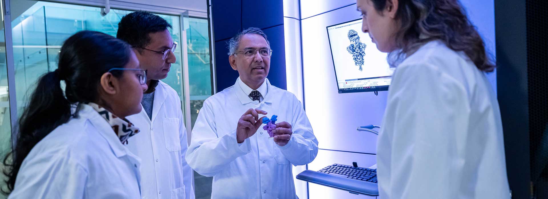
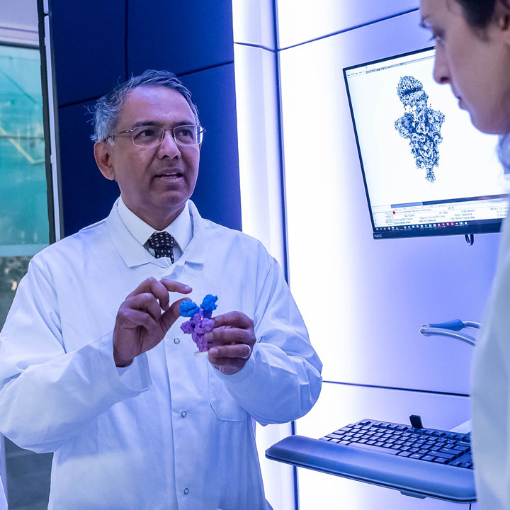
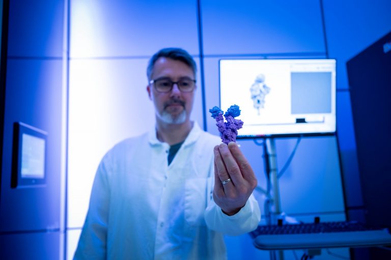
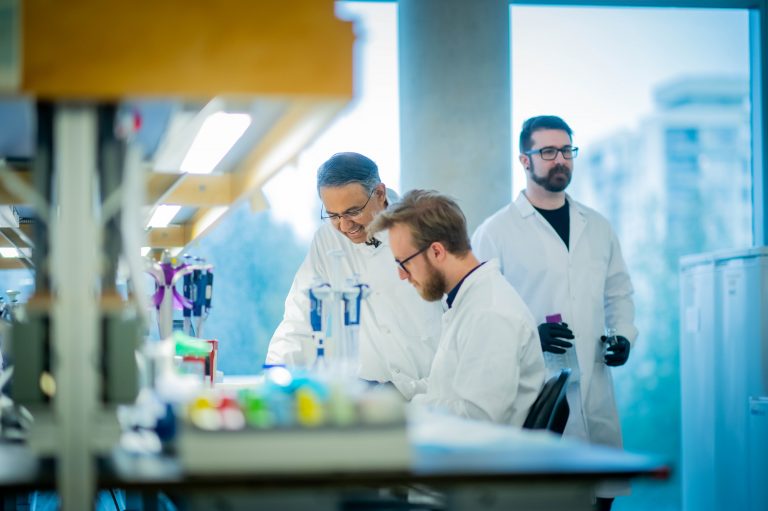
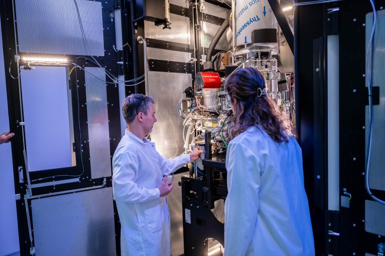
In the heart of Dr. Sriram Subramaniam’s UBC lab, high-powered microscopes as tall as a city bus are allowing researchers to see the very atoms that make up the proteins in our cells — as well as the diseases that affect them.
These cryo-electron microscopes represent a new frontier in drug discovery and development. By firing beams of electrons at samples frozen to nearly -196°C, Dr. Subramaniam and his team are producing detailed structural images of the proteins within human tissue, tumours, bacteria and viruses. With these molecular maps as a guide, the researchers are designing precision treatments tailored to the atomic structure of diseases like cancer, COVID-19, HIV and beyond.
We spoke with Dr. Subramaniam, a professor in the faculty of medicine’s department of biochemistry and molecular biology, and Canada Excellence Research Chair in Prevision Cancer Drug Design, about how cryo-electron microscopy (cryo-EM) is accelerating drug development and helping to shape the future of human health.
What is cryo-EM?
Cryo-EM is a cutting-edge imaging technique that allows scientists to produce detailed images of proteins, the building blocks of our cells. Many of these proteins are thousands of times smaller than the width of a human hair.
With these state-of-the-art microscopes — some of which are up to 12 feet tall — beams of electrons are fired at the speed of light through samples cooled to liquid nitrogen temperatures (-196°C). A camera then captures the electrons to form an image.
Ultimately, hundreds of thousands of these images are taken from proteins oriented in different angles to create an accurate model of protein structures in action at the atomic level.
How is cryo-EM transforming drug discovery and development?
Cryo-EM is already revolutionizing drug discovery for everything from cancer to infectious diseases.
With this technology, we’re able to gain unprecedented insights into the structures of proteins implicated in disease with atomic-level accuracy.
Think of these detailed images as powerful blueprints for designing drugs and vaccines that can specifically target and map precisely to the desired sites on protein surfaces.
What health challenges are you applying this technology to in your lab?
In my lab, we are applying cryo-EM to bring about discoveries in everything from cancer to neuroscience and infectious diseases, such as malaria. When the pandemic hit, we pivoted to help understand, prevent and treat COVID-19, publishing the first atomic-level analysis of spike proteins from most variants of concern including the B.1.1.7 (Alpha) and Omicron variants, revealing how the virus attaches to and infects human cells.
Today, my team at UBC, and at my UBC spin-off company, Gandeeva Therapeutics, are working to accelerate the development of new precision medicines by integrating cryo-EM with AI.
We are particularly interested in taking a cryo-EM-driven approach to the design of molecular glues — the tiny places where proteins bind together. Many diseases are a result of an alteration between protein-protein communication, and so molecular glues represent a significant opportunity to target formerly “undruggable” targets.
What impact do you foresee cryo-EM having on the future of medicine and health care?
Cryo-EM will help us address a key challenge, which is that a one-size-fits-all approach to treatment is frequently not effective for complex diseases, such as cancer.
Looking ahead, I am reminded of the teachings of renowned Canadian-born physician William Osler, who exhorted the medical profession to not merely attempt treating the disease, but to treat the patient who has the disease. Precision medicine is the future of medicine and healthcare — and cryo-EM will play a fundamental role.
Published: February 26, 2024